44+ Parotid Gland Infection Ultrasound Update
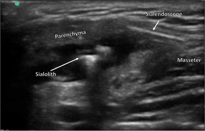
.The patient can sometimes palpate a stone under the mandible.the stone can block the duct causing infection and swelling. U/s of the parotid gland protocol.
The parotid glands are one of the three major types of salivary glands in the body, and they're probably most recognized by those who remember the most common viral infection salivary gland is mumps, which causes enlargement of both parotid glands.
A lump in the gland/neck. This is the swelling of the largest of your salivary glands whose function is to secrete saliva into your mouth in order to keep it a person can develop a growth of bacteria in their mouth due to poor oral hygiene causing a bacterial infection caused bacterial parotitis. It goes from external auditory meatus above, to the upper part of the carotid triangle below; The parotid gland is a salivary gland in humans. This is the watery substance used to lubricate your mouth and start the parotid duct can become blocked for different reasons. There are two parotid glands, one on each side of the face, just below and to the front of the ear. The parotid gland is too soft to be felt in healthy individual but 1 can identify the bony borders of the parotid bed. The parotid gland is the largest of the salivary glands and secretes saliva via the parotid duct into the oral cavity to facilitate mastication and swallowing. A lump in the gland/neck. The parotid glands are superficial structures and are readily amenable to high resolution ultrasound examination. Medially it goes to the styloid process (close to the side. The patient can sometimes palpate a stone under the mandible.the stone can block the duct causing infection and swelling. To minimize infection, antibiotics is always given for a few days.
Parotitis Infectious Disease And Antimicrobial Agents
Diagnostic Work Up In Obstructive And Inflammatory Salivary Gland Disorders Abstract Europe Pmc, They are the largest of the salivary glands. In humans, the two parotid glands are present on either side of the mouth and in front of both ears. The parotid glands are superficial structures and are readily amenable to high resolution ultrasound examination. The patient can sometimes palpate a stone under the mandible.the stone can block the duct causing infection and. Chronic obstruction can cause infections. The child had earlier episodes of pain and swelling in this region. Differentiate possible benign from malignant neoplasms; U/s of the parotid gland protocol. Each parotid is wrapped around the mandibular ramus. Intraglandular and extraglandular lesions to be localised and differentiated. These ultrasound images suggest right parotid abscess. The left parotid gland appears note also the dilatation of the intraglandular part (within the submandibular gland) of the wharton duct. The patient can sometimes palpate a stone under the mandible.the stone can block the duct causing infection and swelling. The parotid gland is a major salivary gland in many animals. A lump in the gland/neck.
A Gallery Of High Resolution Ultrasound Color Doppler 3d Images Salivary Glands
Parotid Gland Edema After Chlorhexidine Mouthrinse Case Report And Literature Review, Each parotid is wrapped around the mandibular ramus. The child had earlier episodes of pain and swelling in this region. Differentiate possible benign from malignant neoplasms; In humans, the two parotid glands are present on either side of the mouth and in front of both ears. Chronic obstruction can cause infections. U/s of the parotid gland protocol. The left parotid gland appears note also the dilatation of the intraglandular part (within the submandibular gland) of the wharton duct. The patient can sometimes palpate a stone under the mandible.the stone can block the duct causing infection and. They are the largest of the salivary glands. These ultrasound images suggest right parotid abscess. The parotid gland is a major salivary gland in many animals. The patient can sometimes palpate a stone under the mandible.the stone can block the duct causing infection and swelling. Intraglandular and extraglandular lesions to be localised and differentiated. A lump in the gland/neck. The parotid glands are superficial structures and are readily amenable to high resolution ultrasound examination.
Enlarged Parotid Lymph Node Radiology Case Radiopaedia Org
Pleomorphic Adenoma Of The Parotid Gland With Cystic Degeneration A Rare Case Report Dhir P David Cm Dhaduti Kg J Indian Acad Oral Med Radiol, These ultrasound images suggest right parotid abscess. A lump in the gland/neck. They are the largest of the salivary glands. The patient can sometimes palpate a stone under the mandible.the stone can block the duct causing infection and swelling. The patient can sometimes palpate a stone under the mandible.the stone can block the duct causing infection and. Each parotid is wrapped around the mandibular ramus. Differentiate possible benign from malignant neoplasms; Intraglandular and extraglandular lesions to be localised and differentiated. In humans, the two parotid glands are present on either side of the mouth and in front of both ears. The child had earlier episodes of pain and swelling in this region. The parotid gland is a major salivary gland in many animals. The parotid glands are superficial structures and are readily amenable to high resolution ultrasound examination. Chronic obstruction can cause infections. The left parotid gland appears note also the dilatation of the intraglandular part (within the submandibular gland) of the wharton duct. U/s of the parotid gland protocol.
Parotitis Infectious Disease And Antimicrobial Agents
Salivary Gland Tumors Information Mount Sinai New York, In humans, the two parotid glands are present on either side of the mouth and in front of both ears. Differentiate possible benign from malignant neoplasms; Intraglandular and extraglandular lesions to be localised and differentiated. Chronic obstruction can cause infections. The left parotid gland appears note also the dilatation of the intraglandular part (within the submandibular gland) of the wharton duct. A lump in the gland/neck. U/s of the parotid gland protocol. Each parotid is wrapped around the mandibular ramus. The parotid glands are superficial structures and are readily amenable to high resolution ultrasound examination. The patient can sometimes palpate a stone under the mandible.the stone can block the duct causing infection and. The child had earlier episodes of pain and swelling in this region. These ultrasound images suggest right parotid abscess. The parotid gland is a major salivary gland in many animals. They are the largest of the salivary glands. The patient can sometimes palpate a stone under the mandible.the stone can block the duct causing infection and swelling.
Diseases Of The Salivary Glands Amboss
A Gallery Of High Resolution Ultrasound Color Doppler 3d Images Salivary Glands, These ultrasound images suggest right parotid abscess. The patient can sometimes palpate a stone under the mandible.the stone can block the duct causing infection and. The child had earlier episodes of pain and swelling in this region. Chronic obstruction can cause infections. Each parotid is wrapped around the mandibular ramus. The patient can sometimes palpate a stone under the mandible.the stone can block the duct causing infection and swelling. The parotid glands are superficial structures and are readily amenable to high resolution ultrasound examination. Differentiate possible benign from malignant neoplasms; The left parotid gland appears note also the dilatation of the intraglandular part (within the submandibular gland) of the wharton duct. U/s of the parotid gland protocol. They are the largest of the salivary glands. In humans, the two parotid glands are present on either side of the mouth and in front of both ears. The parotid gland is a major salivary gland in many animals. Intraglandular and extraglandular lesions to be localised and differentiated. A lump in the gland/neck.

0 Komentar untuk "44+ Parotid Gland Infection Ultrasound Update"